


How Does an XRF Spectrometer Work?
X-ray fluorescence (XRF) spectrometry is the measurement and analysis of samples excited by incident radiation. It is a non-destructive method of material characterization, allowing for accurate investigations into the elemental and chemical make-up of metals, ceramics, geological samples, historical items, and more. This method relies on an XRF spectrometer, and specialized sample preparation routines to ensure excellent measurement accuracy and repeatability.
The two primary components of an XRF spectrometer are an x-ray output and a sensitive detector capable of determining fluorescent x-rays from the incident light. This array emits a beam of x-rays or gamma rays into a sample, exciting electrons within the sample’s inner atoms and displacing them from their orbital shells. Atoms replace the vacancies left by these electrons by bringing down electrons from higher orbital shells – losing the increased binding energy associated with outer orbits in the process. This release of energy is registered as fluorescence, a phenomenon that is unique to each element. XRF spectrometers can register this rapid bloom of energy in real-time – but they require samples to be specifically prepared to ensure that results can be accurately analyzed.
There are numerous methods of sample preparation for analysis by an XRF spectrometer, including:
- Solid sample preparation;
- Liquids;
- Powders;
- Pellets;
- Fused beads.
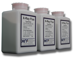
Solid samples can be analyzed by an XRF spectrometer with little to no sample preparation, but surface area variations and irregularities can introduce a high degree of error analysis due to the sensitive calibration of the XRF equipment. Ideally, solid samples will be cut and finished using grinding methods and an application of a fusion flux mixture. Granular mixtures of 64.7% lithium metaborate and 35.3% lithium tetraborate fusion flux are ideal for solid sample preparation, with optimal analytical results for various aluminosilicates.
Liquid samples are prepared for XRF analysis through the application of a support film which exhibits little interference with the XRF spectrometer’s incident beam. Powders are prepared using a similar methodology.
Pellets provide better insights into the homogenous composition of a sample. They are produced specifically for analysis in an XRF spectrometer by grinding a sample to a fine powder using a laboratory crusher or pulverizer and mixing it with a binding agent, prior to compressing the mixture into a denser sample. This provides exceptional analysis quality; however, pellets retain mineralogical structures which interfere with or alter the fluorescent process, impacting results accuracy.
The preparation of samples as fused beads or discs reduces this issue, providing a near-perfect representation of the homogenous XRF reaction. This process involves crushing the sample, then mixing it with a fusion flux of varying ratios into a platinum crucible. The mixture is then heated to temperatures up to 1000°C (1832°F) and cast as a disc or a bead. An XRF spectrometer can accurately examine a fusion bead or disc sample due to the reduction of its mineralogical matrixes.
XRF Preparation from XRF Scientific
XRF Scientific is a global manufacturer and supplier of laboratory equipment and fusion flux mixtures for varied sample preparation methodologies. We provide a catalog of products which can optimize sample preparation for analysis by an XRF spectrometer, improving results accuracy and laboratory throughput.
If you would like any more information about preparing samples for XRF analysis, or supporting XRF spectrometer applications with additional equipment, please do not hesitate to contact us.